


Suffering from severe internal diseases, malnutrition and aging, the growth of nails will slow down and their structure will also change. Only a doctor can accurately determine the cause of the violation based on the results of the examination and microscopic examination.
But to understand what happens to the nails of the legs or hands, you can use the photos for different types of fungal diseases.
Causes of nail deformation
Nail infections (onychomycosis) caused by molds, yeast-like fungi and dermatophytes show similar symptoms.
All types of toenail or toenail fungus deform the nail plate, changing its transparency, gloss, and color. This color can be seen in the photos shown.
Nail changes not only occur in onychomycosis, but also in injuries, chronic paronychia (inflammation of nail folds), psoriasis, hand eczema, dermatitis. Before determining the presence of a fungal infection, you need to consider all possible options.
Signs of fungal infection
The most significant signs of fungal infection are changes in the color of the nail plate, the presence of nail separation, surface changes-transverse, longitudinal grooves, depressions, thickening, nail destruction on the nail plate.
The pink color of healthy nails is determined by the transparency of the nail plate and the blood vessels visible through it. Onychomycosis will make the nails lose their transparency, and their color will turn brown, yellow, and rarely green and black.
Candida and dermatophytes can cause onycholysis-separation of the affected part of the nail. When infected with dermatophytes, onychomycosis was observed from the distal end of the nail, and when infected with Candida, the nail stayed behind the nail bed at the bottom of the crescent moon area.
The symptom of Candida may be inflammation of the outer lip-paronychia. The disease has a bacterial form caused by Streptococcus and Staphylococcus and non-infectious bacteria-eczema, psoriasis, systemic vasculitis.
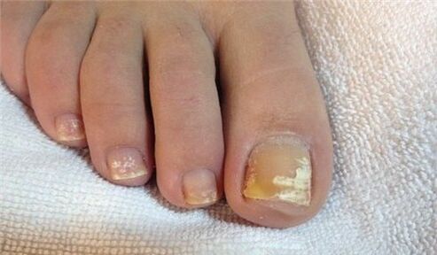
When the toenail is affected by the rubrum fungus, the plate will also be affected, as you can see in the photo, the roller will not be affected by the infection. The plate turns yellow and thickens strongly, and the accumulated fungal clumps can be clearly distinguished under the plate.
Nail fungus due to skin fungal infection
95% of all nail fungus cases are caused by dermatophytes Trichophyton and Mentophyton.
Red Trichophyton Infection
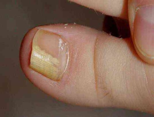
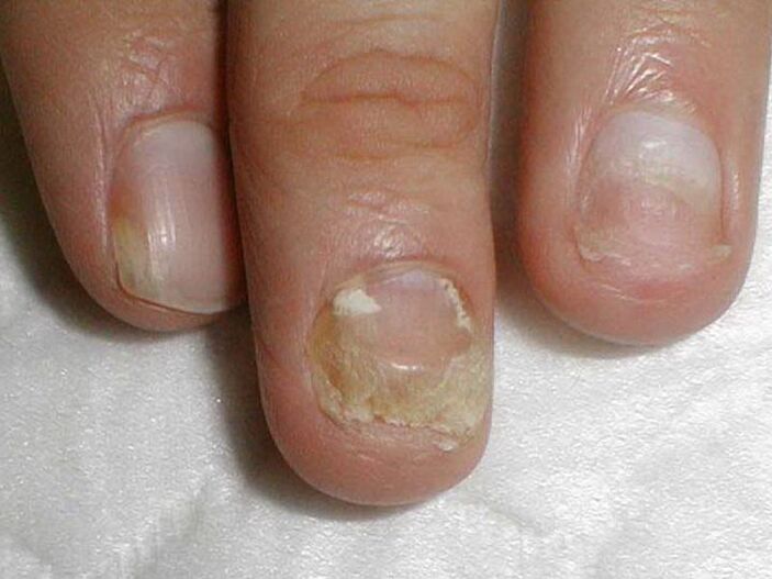
Onychomycosis begins when the fungus penetrates under the nail plate from the free edge. Fungal infections are indicated by the appearance of yellowish spots, the area of which is an uneven, fragile surface of the distal (distal) edge of the nail.
Thedistal-lateral formof fungal skin infections of Rhodophyton is very common. In the photo, you can see that the stain caused by the introduction of fungus is located outside the periphery of the nail.
Usually, red trichophyton can affect the big toe, causing hyperkeratosis-the fungus builds up between the nail plate and the nail bed, which looks like scattered light yellow clumps.
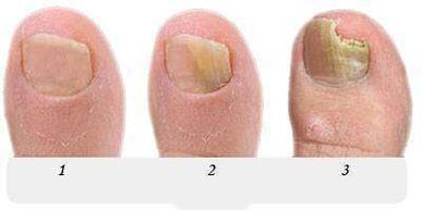
At this stage, the fungus occupies a very small part of the nail, as shown in the photo presented, with the help of topical treatment, it is possible to cope with the initial attack of onychomycosis.
Without treatment, the stain will grow and gradually affect the entire edge of the nail, and then move to half a month. In the photo, the nail fungus looks like a yellowish streak, pointing towards the growth area of the nail plate.
Distal form of nail fungus(usually found on the big toe), the distal edge of the nail may have a yellowish infection spot in the center. See it in the photo.
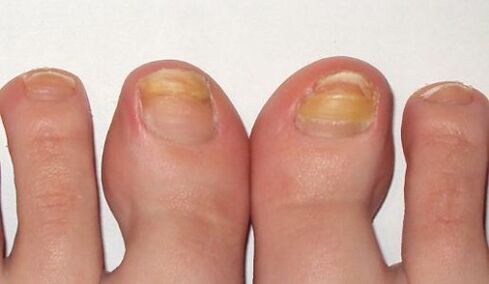
In the late stage of leg fungus, as shown in the photo, several nails were affected, and treatment was no longer limited to local drugs and pills. In addition to antifungal drugs, nail hardware must be cleaned to partially or completely remove the nail plate.
As shown in the picture, all known antifungal agents should be used for long-term treatment and hyperkeratosis of the legs caused by Trichophyton vulgaris should be treated.
The fungal infection is accompanied by total nail damage spreading to the entire area of the nail plate, and the nail is completely destroyed.
Another representative of dermatophytes, Trichophyton mentagrophytes, can also cause complete fungal infection of the nails.
Trichophyton infection
After thoroughly destroying the toenails with Trichophyton fungus, the nail plate was deformed. The photo showed that the nail plate became thicker, changed its structure, collapsed, and light yellow spots appeared on the entire surface.
Infection of nails with this dermatophyte usually causes superficial white onychomycosis of the big toe, but rarely causes white ringworm of the small toe.
This fungus does not actually occur on the nails of the hands. It often causes interdigital dermatophytosis on the legs, as shown in the photo, and requires simultaneous treatment of the skin of the feet and nails.
The symptoms of nail fungal infection are usually on the feet, which are white spots of various sizes, as shown in the photo, which are reminiscent of diphtheria-a disease of the nail plate itself.
But unlike diphtheria, diphtheria is a leukoplakia caused by bubbles in the nail layer, which is the result of the activity of Trichophyton in fungal infections.
Rarely, superficial white onychomycosis is caused by a mold; in AIDS, the pathogen of this fungus can be Rhodophyton, and affects the nails of the feet and hands.
Nail changes due to Candida infection
Fungus usually occurs in women, it affects the nails of working hands, and working hands are often in contact with water.
For candidal onychomycosis, it is characterized by a proximal form of infection, in which the fungus first affects the nail crease at the fingernail part, and then penetrates into the growth zone and nail bed. Then it gradually moved along the nail from the bottom to the edge, capturing a larger nail area.
The cause of Candida onychomycosis is Candida albicans. As you can see in the picture, this fungus invades toenails and nails, spreading from the meniscus at the bottom of the nail plate to the free edge.
Signs of Candida nail infectionCandida albicans is a nail fold (paronychia), the cuticle is separated from the nail plate, painful, and the pus is discharged when the bacterial infection is attached.
Candida albicans can penetrate the nail from the free edge of the nail. In this case, they are talking about remote infections, usually combined with cutaneous candidiasis.
 As shown in the picture, the treatment of Candida on the nails of the hands and feet damages more than half of the nail plate area. It includes not only measures against onychomycosis, but also measures to reduce onychomycosis. The activity of Candida in the natural reservoir where it is stored-intestines, oral cavity, genital mucosa. . .
Mold infestation
Molds cause fungi less frequently than Candida or dermatophytes. As shown in the photo, the main symptom of toenail infection with mold is to change the color of the nail plate to blue, black and green in.
Signs of toenail mold may be black spots, spots on the nail plate, or black longitudinal stripes (as shown in the picture).
Fungal preparations
Antifungal agents containing fluconazole, ketoconazole, terbinafine, itraconazole, griseofulvin are used to treat nail fungus caused by dermatophytes, such as this picture.
Antifungal agents containing terbinafine are effective against fungal skin infections.
The antifungal agent of voriconazole has high activity against dermatophytes.
forandfor the treatment of feet, hands and nail mold against Candida yeaston. The scope of action includes molds such as Aspergillus, Fusarium, and Penicillium.
Itraconazole-based formulations can deal with mold.
Fungal nail disease
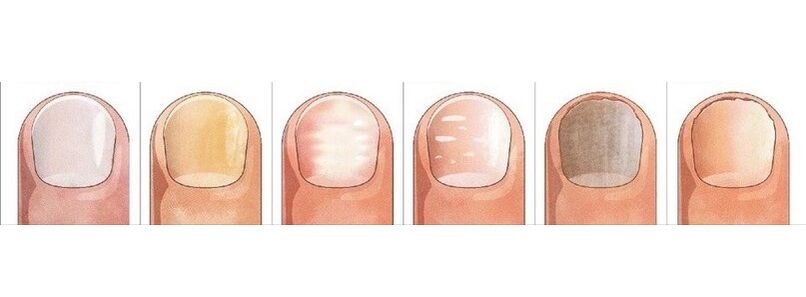
Nailssometimes appear light graywith eczema. In this case, the nail plate will move away from the nail bed, which is observed with fungi.
is very similar to psoriasisPsoriasis is very similar. For this disease, not only will thecolor change, but also thenail plate will become thicker.
Found depressions on its surface and noticed the separation of the nail plate from the nail bed. But there is a difference with fungi: in psoriasis, over time, the free and healthy parts of the toenails are separated by pink and yellowed bands.
light blueMake nailsPseudomonas nail infection. Frequent mechanical friction of nail creases can cause surface grooves and nail undulations.
Diphtheria leukoplakiaThe appearance ofrelated to metabolic disorders may also be mistaken for superficial white fungus with larger spots.
Changes in the color and shape of the nail that caused the injury. The big toe is in greatest danger. Both nails and fungus can be injured and thickened and darkened.

The difference between injury and fungus is that the changes in the injury process are only visible on the injured finger, the nails on other fingers remain unchanged, and will not be infected from the diseased finger like onychomycosis.
The consequence of trauma may be that the nail is partly separated from the nail bed, forming a cavity, and under adverse conditions, the fungus will quickly colonize it.
Under the influence of iron deficiency anemia and hormonal diseases, the nail plate can be separated from the nail bed under the action of light (photolysis). Splitting, nail shedding occurs along with lichen erythema, bullous skin disease, and nail trauma.
However, you can finally make sure that the conclusion is correct and start treatment. You can only do this after you seek the help of a dermatologist-dermatologist or mycologist-physician who treats mycosis.